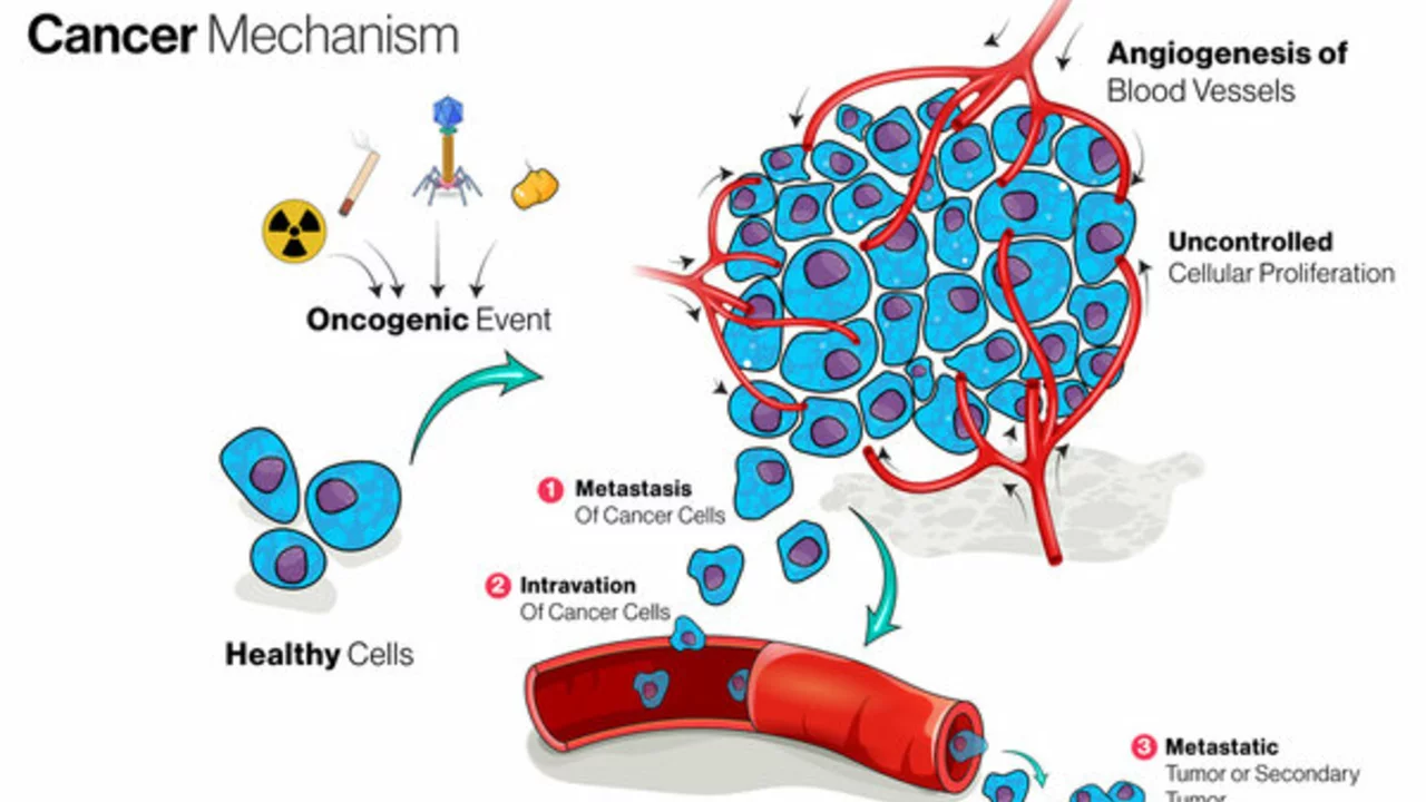
Understanding Tumor Basics
First things first, let's start with the basics. What is a tumor? And no, I'm not referring to the constantly shedding ball of fur and energy known as Dexter, my lovable Cocker Spaniel. In medical terms, a tumor (or neoplasm if you want to impress at the next dinner party) is an abnormal mass of tissue that forms when cells grow and divide more than they should or when they don't die when they should.
Fascinating, isn't it? The wonders of nature! Just think about it for a second. It's like your body, in all its wisdom, decides to throw a wild cell party with no closing time in sight. And the result? A mass of rogue cells gate-crashing your body's normal functions like an unwelcome house guest. Kind of like Dexter when I try to take a nap.
Breaking Down Growth Rates
Moving on, let's talk growth rates. Just like that weed in your garden that grew to the size of a small tree in a week (seriously, where did that thing come from?), tumors are known for their rapid growth rates. But how fast do they actually grow?
Well, it turns out it depends on a few things. Much like Dexter, who will do anything to get ahold of his neighbors chew toy, tumor growth can be aggressive. But unlike Dexter, who eventually gets tired and takes a nap, tumors rarely show signs of slowing down. Though they do occasionally pause for a rest, the characteristics of the tumor, such as what type it is, and the overall health and age of the patient are all key factors in determining how fast a tumor can grow.
Different Strokes for Different Folks
Now if you thought all tumors were created equal, think again. No, siree! Just like people and pets (you don't see Dexter chasing his tail now, do you?), every tumor is unique. Some grow slowly and steadily, ticking off the days and nights with a relentless regularity. On the other hand, some tumors, are the party-poopers, growing rapidly, spreading and causing havoc faster than you can say "Dexter, drop it!".
So what's driving these differences? Simply put, it's the type of cell the tumor originates from, its location in the body, and the unique genetic characteristics of the tumor itself. And yes, you're right. It does sound a bit like a James Bond villain strategy. But in reality, it's a complex process, with each factor interplaying and influencing the other in often unpredictable ways. Kind of like throwing a ball for Dexter, expecting him to fetch it, and instead, he returns with the neighbor’s newspaper.
Keeping An Eye On The Ball
Finally, let's talk about tracking tumor growth. Yes, there's actually a way to keep an eye on these sneaky customers! The studies and scans doctors conduct provide them with useful information on the size, spread, and characteristic of the tumor. I had a similar experience too. I once had a growth on my neck, turned out to be benign thankfully. And let me tell you, the process of tracking its size and shape was much like watching Dexter grow from a pint-sized pup to the burly rambunctious canine he is today.
Seriously though, medical professionals utilize various techniques – CT scans, MRIs, ultrasounds, to monitor tumor growth. You might be wondering, 'Arvid, how does that work?’ Interestingly, these techniques work similarly to how I monitor Dexter’s behavior. One minute he's calm and poised, the next, he's zipping around the house like a pint-sized hurricane. With the medical scans, the doctors use the images captured to measure the size, direction and pace of the tumor’s growth, allowing them to lay out a plan of attack.
There you have it folks! The lowdown on tumor growth rates. So, here’s a piece of advice. Keep an eye out for changes in your body, just as you would on your pet's behavior. Early detection can make all the difference, be it with humans or pets. Stay proactive, and remember to take care of your health. And now, I'm off to wrestle with Dexter who has very strategically plopped himself on my laptop. Bless his little cotton socks!


Understanding the nuances of tumor proliferation enables clinicians to tailor surveillance protocols, thereby optimizing patient outcomes. By integrating serial imaging metrics with individualized risk assessments, we can anticipate growth trajectories and intervene with greater precision.
When you consider the biology behind exponential tumor expansion, it becomes clear that not all neoplasms follow a single kinetic pattern; some adhere to a Gompertzian curve while others display a more linear progression. Imaging modalities such as CT and MRI provide volumetric data that, when plotted over time, reveal the underlying growth constant. Armed with that knowledge, clinicians can schedule follow‑up scans at intervals that balance early detection with patient convenience. Moreover, incorporating functional imaging like PET can highlight metabolic activity, offering an extra layer of insight beyond mere size. Ultimately, a multidisciplinary approach-combining radiology, pathology, and oncology-ensures that treatment plans remain both proactive and adaptable.
I appreciate the concise overview and will keep the timing of follow‑up scans in mind for my patients.
Honestly this kinda sounds like a hype train, but ya gotta admit the hype is legit cuz tumors don’t wait for anyone’s schedule, they just keep pushin forward.
When we talk about tumor growth we are really diving into a complex interplay of cellular replication and microenvironmental pressures that shape how quickly a mass can expand over time. The rate is not a simple constant it fluctuates based on genetic mutations the availability of nutrients the immune response and even the mechanical constraints of surrounding tissues. Each of these factors can accelerate or decelerate the expansion in ways that are often unpredictable. The classic models like exponential or Gompertzian curves provide a framework but the real world rarely fits neatly into a single equation. Clinicians therefore rely on sequential imaging to capture real data points that reflect the true behavior of a particular lesion over weeks or months. By comparing diameter measurements or volumetric assessments they can calculate a specific growth rate for that tumor that informs prognosis and treatment planning. The importance of early detection cannot be overstated because catching a fast‑growing lesion before it breaches critical structures can make the difference between curative surgery and palliative care. Moreover advancements in functional imaging allow us to see not just size changes but also metabolic activity which adds another layer to our understanding of aggressiveness. In a practical sense this means that a patient with a small but metabolically active tumor may be treated more aggressively than one with a larger but indolent mass. The takeaway is that tumor growth is a dynamic process that must be monitored closely and personalized strategies are essential for optimal outcomes. Serial scans also help differentiate treatment‑induced necrosis from true tumor progression, reducing the risk of premature therapy changes. Additionally, molecular profiling can reveal proliferative markers such as Ki‑67, providing a histologic correlate to imaging‑based growth estimates. Combining these modalities creates a comprehensive picture that guides multidisciplinary decision‑making. Patients benefit from transparent communication about expected growth patterns, which can alleviate anxiety and improve adherence to monitoring schedules. Ultimately, a proactive surveillance strategy empowers clinicians to intervene at the optimal window, potentially improving survival and quality of life.