Mycosis Fungoides Staging & Treatment Guide
Enter disease stage and age group to see recommended treatment approach.
| Modality | Typical Stage | Response Rate | Common Side Effects |
|---|---|---|---|
| Topical corticosteroids | IA/IB | 30-50% | Skin thinning, stretch marks |
| PUVA phototherapy | IA/IIA | 55-70% | Sunburn-like redness, nausea |
| Extracorporeal photopheresis (ECP) | IIB/III | 40-60% | Low blood pressure, fatigue |
| Brentuximab vedotin | III/IV | 70-80% | Neuropathy, neutropenia |
Quick Takeaways
- Mycosis Fungoides is the most common form of cutaneous T‑cell lymphoma, starting as skin patches and possibly progressing to tumors.
- Early signs include persistent, itchy patches that don’t respond to usual creams.
- Diagnosis relies on a skin biopsy, immunophenotyping, and staging studies.
- Treatment ranges from topical steroids to phototherapy and targeted systemic drugs, chosen based on stage and patient health.
- Remission is achievable for many patients; regular follow‑up and skin monitoring are key.
Understanding Mycosis Fungoides
When you first hear the name Mycosis Fungoides is a rare, slow‑growing cancer of the skin’s T‑cells, it can feel like a wall of medical jargon. In plain terms, it’s a type of skin lymphoma that begins as red, scaly patches and can, over years, develop into thicker plaques or even tumors. Though it accounts for less than 1% of all non‑Hodgkin lymphomas, it is the most common variant of cutaneous T‑cell lymphoma (CTCL), a group of cancers that originate in the immune cells residing in the skin.
What makes Mycosis Fungoides tricky is its chameleon‑like presentation. Early lesions often look like eczema, psoriasis, or fungal infections, which is why patients can wander through months of misdiagnoses before getting the right answer.
Spotting the Early Signs
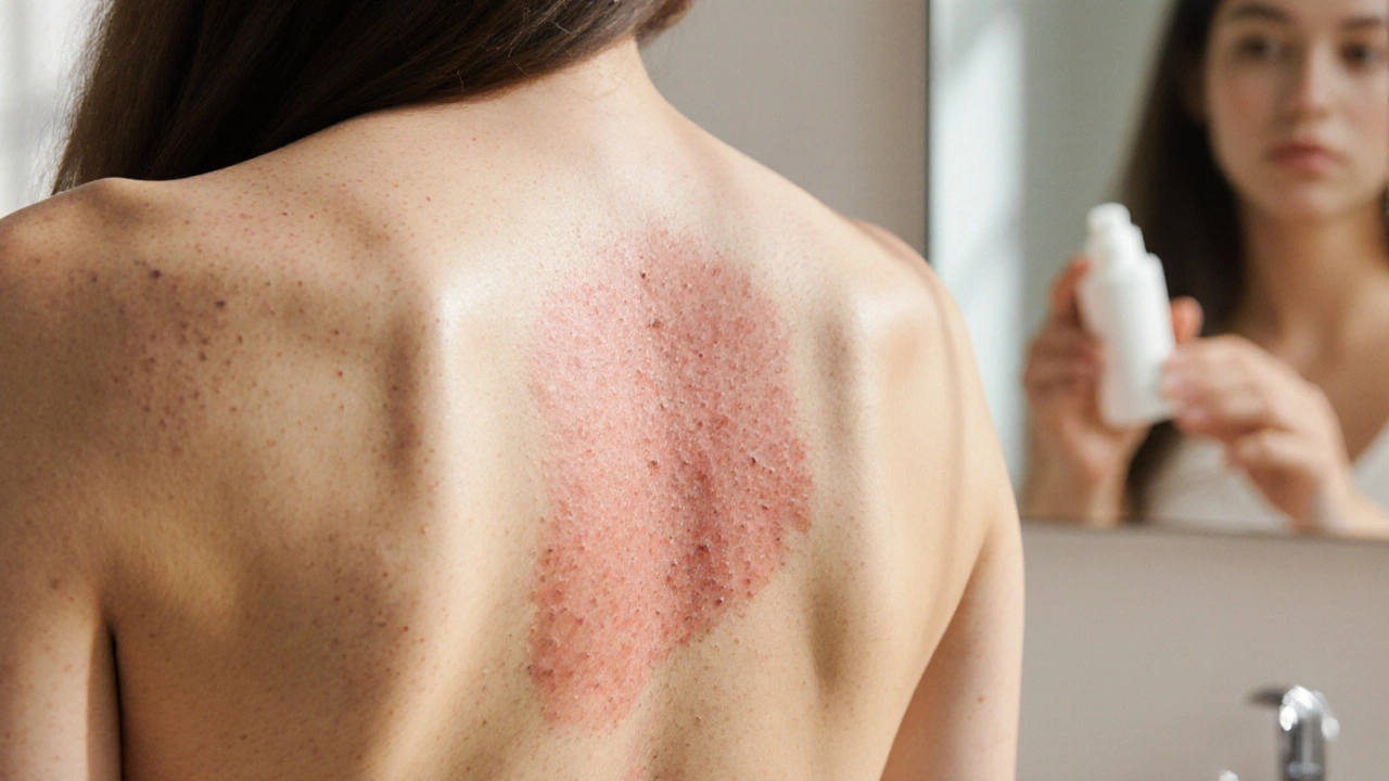
Most patients notice a persistent rash that itches or burns, usually on the torso, buttocks, or groin. The patches are typically:
- Slow‑growing, lasting weeks to months.
- Flat, pink‑to‑red, sometimes with a fine scale.
- Unresponsive to over‑the‑counter steroids or antifungals.
- Accompanied by subtle skin thinning in later stages.
If you’ve tried a standard skin biopsy for another condition and still have lingering patches, it’s a good cue to ask your dermatologist about a deeper investigation specifically for CTCL.

The Diagnostic Roadmap
Getting a firm diagnosis usually involves three steps:
- Skin biopsy: A small piece of the lesion is removed under local anesthesia. Pathologists look for atypical T‑cells that express markers like CD4 and loss of CD7.
- Immunophenotyping: Using flow cytometry or immunohistochemistry, doctors confirm the T‑cell lineage and rule out other lymphomas.
- Staging work‑up: Depending on the findings, imaging (CT or PETscan) and blood tests assess whether the disease is limited to the skin (early stage) or has spread to lymph nodes, blood, or internal organs (advanced stage).
Staging follows the TNMB system (Tumor, Node, Metastasis, Blood). Early stages (IA‑IIA) involve only patches or plaques, while stages IIB‑IV indicate tumors, nodal involvement, or blood disease. Knowing your stage guides the treatment plan.
Choosing the Right Treatment
Therapy is highly individualized. Doctors weigh the disease stage, patient age, skin condition, and personal preferences. Below is a snapshot of the most common options.
| Modality | Administration | Typical Stage | Response Rate | Common Side Effects |
|---|---|---|---|---|
| Topical corticosteroids | Cream/ointment applied daily | IA‑IB (patches/plaques) | 30‑50% | Skin thinning, stretch marks |
| PUVA phototherapy | Oral psoralen + UVA sessions 2‑3×week | IA‑IIA | 55‑70% | Sunburn‑like redness, nausea, long‑term skin aging |
| Extracorporeal photopheresis (ECP) | Blood processed outside body, UV‑A exposure | IIB‑III (tumors or blood involvement) | 40‑60% | Low blood pressure, fatigue |
| Brentuximab vedotin | IV infusion every 3weeks | III‑IV (advanced disease) | 70‑80% | Peripheral neuropathy, neutropenia |
Here’s how doctors usually pick:
- Early stage (IA‑IB): Topical steroids or mild phototherapy are first‑line because they’re low‑risk.
- Intermediate stage (IIA‑IIB): Narrow‑band UVB or PUVA become the go‑to, sometimes combined with low‑dose oral retinoids.
- Advanced stage (III‑IV): Systemic agents like brentuximab vedotin or interferon‑α, often alongside ECP, are considered.
Living Through Treatment
Treatment isn’t just a medical checklist; it’s a daily reality. Patients often report:
- Fatigue from frequent clinic visits.
- Skin sensitivity that makes everyday activities uncomfortable.
- Emotional ups and downs as visible lesions improve or flare.
Practical tips can make a big difference:
- Moisturize aggressively: Fragrance‑free creams keep skin barrier intact.
- Sun protection: Even on non‑phototherapy days, SPF30+ reduces cumulative UV damage.
- Support network: Joining a Mycosis Fungoides patient group (online or in‑person) provides shared coping strategies.

Reaching Remission
Remission means the skin lesions have cleared or stabilized to a point where they no longer progress. It can be:
- Complete remission: No visible disease for at least 6months.
- Partial remission: Lesions shrink but some patches remain.
Key cues for remission include:
- Absence of new lesions for three consecutive visits.
- Biopsy of previously involved skin showing no atypical T‑cells.
- Stable blood counts and imaging, if previously abnormal.
Even after remission, lifelong surveillance is essential. Dermatology visits every 3‑6months, periodic skin biopsies of any suspicious area, and blood work for atypical lymphocytes keep the disease in check.
A Patient’s Story (Illustrative)
Emily, a 42‑year‑old teacher, first noticed a stubborn rash on her lower back at age 38. After trying several over‑the‑counter creams, the patch kept expanding. A dermatologist performed a skin biopsy, and the pathology report confirmed Mycosis Fungoides, stage IB.
Her treatment plan began with potent topical steroids for three months, followed by PUVA phototherapy three times a week. Within six months, the rash faded dramatically, but she experienced occasional nausea from the oral psoralen. Adjusting her diet and taking anti‑nausea medication helped.
Two years later, a routine skin check revealed a small plaque on her thigh. Early‑stage ECP was added, and the plaque cleared in three cycles. Today, Emily enjoys a stable partial remission, attends quarterly dermatologist appointments, and volunteers with a local CTCL awareness group.
Emily’s journey underscores two points: early detection and a flexible, stage‑adapted treatment strategy can turn a frightening diagnosis into a manageable chronic condition.
Next Steps Checklist
- Schedule a full‑body skin examination if you have persistent, unresponsive patches.
- Ask your dermatologist about a skin biopsy and immunophenotyping for accurate staging.
- Discuss treatment goals: symptom control, disease clearance, or long‑term remission.
- Consider joining a support community for emotional guidance.
- Plan regular follow‑up visits even after remission to catch early signs of recurrence.
Frequently Asked Questions
What exactly is Mycosis Fungoides?
Mycosis Fungoides is the most common form of cutaneous T‑cell lymphoma, a cancer that starts in the skin‑resident immune cells. It typically begins as red, scaly patches that can evolve into thicker plaques or tumors over years.
How is the diagnosis confirmed?
Diagnosis relies on a skin biopsy examined for atypical T‑cells, followed by immunophenotyping to confirm the CTCL profile. Staging tests such as CT scans or blood work determine how far the disease has spread.
When is phototherapy recommended?
Phototherapy, especially PUVA or narrow‑band UVB, is the go‑to for early‑ to intermediate‑stage disease (IA‑IIA). It works by targeting the malignant T‑cells in the skin while sparing deeper tissues.
Can Mycosis Fungoides be cured?
Complete cure is rare, but many patients achieve long‑lasting remission with modern therapies. Ongoing monitoring is essential because the disease can recur.
What lifestyle changes help during treatment?
Gentle skin care, strict sun protection, balanced nutrition, and stress‑reduction practices improve tolerance to therapy and support skin healing.


Reading through the journey feels like a beacon for anyone stuck in that fog. The step‑by‑step breakdown of staging makes the whole process less scary. I love how the author mixes clinical facts with real‑world tips, it’s the sweet spot between science and hope. Keep the emojis coming 😊, they remind us that resilience can be playful.
The guide pretends to be comprehensive but glosses over the harsh reality of systemic toxicities. It‑like glosses over cost issues that burden patients daily. The language sounds sanitized, as if pharma wrote it. A more brutal honesty would serve the community better.
Early detection saves lives.
While the guide does mention side effects, it also provides actionable mitigation strategies that many patients overlook. The inclusion of moisturization and sun protection isn’t merely fluff, it’s evidence‑based care. 😊
The staging matrix is basically a clinical decision tree, and that algorithmic framing helps clinicians allocate resources efficiently. From a health‑system perspective, the cost‑effectiveness of PUVA versus newer biologics is a crucial factor.
A practical tip many forget is to keep a symptom diary; noting flare‑ups alongside treatment dates can reveal hidden triggers. Also, collaborating with a multidisciplinary team-dermatology, oncology, and nutrition-boosts overall outcomes. Consistency in skin care routines can’t be overstated.
If we glorify decision trees, we risk turning patients into mere data points, forgetting their lived narratives. The jargon‑laden approach can alienate the very folks who need clarity. A dash of humanity in the protocol would make the roadmap feel less sterile. Let’s remember the skin is more than a battleground.
Your diary suggestion is gold; many patients say tracking rash patterns saved weeks of trial‑and‑error. Pairing that with community support groups gives emotional ballast. Keep spreading this practical wisdom!
Mycosis Fungoides, as the most prevalent cutaneous T‑cell lymphoma, demands an interdisciplinary diagnostic pathway. The initial clinical suspicion should arise when a lesion persists beyond eight weeks despite standard eczema therapy. A full‑thickness skin biopsy, stained for CD3, CD4, and loss of CD7, remains the gold standard for histopathological confirmation. Immunophenotyping by flow cytometry further delineates the neoplastic clone and excludes peripheral T‑cell lymphoma. Staging according to the TNMB system integrates tumor burden, nodal involvement, visceral spread, and blood involvement, each dictating therapeutic intensity. Early‑stage IA‑IB disease often responds to high‑potency topical corticosteroids applied twice daily for four to six weeks, with careful monitoring for skin atrophy. Phototherapy modalities, particularly PUVA and narrow‑band UVB, achieve response rates above fifty percent and should be instituted when topical agents fail. For intermediate disease (IIA‑IIB), combination regimens of low‑dose oral retinoids such as bexarotene with phototherapy have demonstrated synergistic clearance. Advanced stages (III‑IV) merit systemic interventions; brentuximab vedotin, an anti‑CD30 antibody‑drug conjugate, offers response rates approaching eighty percent but necessitates vigilant neuropathy surveillance. Extracorporeal photopheresis, administered on consecutive days bi‑weekly, provides immunomodulatory benefits in erythrodermic presentations and can be combined with interferon‑alpha. Supportive skin care, including fragrance‑free emollients applied immediately after bathing, preserves barrier function and reduces secondary infection risk. Rigorous photoprotective measures, such as broad‑spectrum SPF 30+ sunscreen, are indispensable regardless of therapy type to mitigate cumulative UV damage. Nutritional optimization, emphasizing omega‑3 fatty acids and antioxidant‑rich foods, may enhance treatment tolerability and improve quality of life. Psychological support, through counseling or peer‑led groups, addresses the emotional turbulence that accompanies chronic dermatologic illness. Surveillance protocols recommend dermatologic assessment every three to six months post‑remission, supplemented by targeted biopsies of any suspicious area. Ultimately, a patient‑centered approach, blending evidence‑based modalities with individualized lifestyle adaptations, yields the most favorable long‑term outcomes.
The comprehensive outline aligns with the standards set by leading American oncologic societies, reflecting our nation's commitment to evidence‑based care. However, the guide should emphasize that domestic clinical trials often outperform foreign alternatives in accessibility. By prioritizing home‑grown therapies, we reinforce our healthcare sovereignty. A patriotic endorsement of these protocols would further galvanize patient confidence.
Wow the guide is like a saga of skin battles, every stage feels like a new chapter in a thriller! It keeps you on the edge of your seat.
Totally feel ya, ya kno each new tecnique can feel like another twist in the plot. Keep your head up, and leet's stick togther through the ride. We're all in this story together.
I doubt the pharma narrative behind this guide.